Modern dentistry in Bhopal includes digital X-rays, intra-oral scanners, CBCT imaging, laser-assisted procedures, CAD/CAM and 3D-printed restorations, microscope-assisted endodontics, and digital smile design — all available at established clinics that combine specialist training with current technology.
Medically reviewed by Dr. Saurabh Shrivastava, BDS MDS Prosthodontist, Certified Digital Smile Designer (DSD) (DCI: A-04860). Last updated: May 2026.
Modern dentistry in Bhopal now includes the same core technologies used in leading clinics elsewhere — digital X-rays, intra-oral scanners, CBCT imaging, laser-assisted procedures, CAD/CAM and 3D-printed restorations, microscope-assisted endodontics, and digital smile design — and these are applied alongside specialist training at Smile Gallery Dental Wellness Centre, Arera Colony, by Dr. Saurabh Shrivastava, BDS MDS Prosthodontist (DCI: A-04860), Certified Digital Smile Designer, for patients from Shahpura and across the city. Modern dental treatment outcomes — root canal therapy planned with care over 10 years, implants at 98% survival at 5 years — depend on technology used well, not technology alone.
What Counts as Modern Dentistry in Bhopal Today
In short: modern dentistry in Bhopal is digital, image-guided, and minimally invasive — digital X-rays, intra-oral scanners, CBCT, lasers, CAD/CAM, and microscope-assisted endodontics.
Modern dentistry is digital, image-guided, and minimally invasive where possible. Film X-rays have largely given way to digital sensors. Putty impressions are replaced by intra-oral scans for many cases. CBCT scans provide 3D views for implant and complex root canal planning. Diode and erbium lasers handle selected soft-tissue work. CAD/CAM and 3D printing produce precise crowns, bridges, and aligners faster than older laboratory methods. Microscopes magnify the inside of teeth for precise endodontic work.
How to Identify a Clinic That Uses These Tools Well
Walk through the clinic before booking complex work. Look for digital sensors instead of film X-rays. Look for intra-oral scanners. Check that autoclave sterilisation is in plain view. Ask whether before-and-after photos are from the clinic’s own patients. The answers reveal more than any marketing material.
According to Dr. Saurabh Shrivastava, MDS Prosthodontist: "Modern dentistry in Bhopal is not just about having the equipment — it is about using it correctly at every stage. An intra-oral scanner takes a more accurate impression than putty, but only if the operator knows how to position it and read the scan. Technology reduces human error; it does not replace clinical judgement."
Specialist Training Behind the Technology
Quick answer: the best modern dentistry in Bhopal pairs current technology with specialist training — tools reduce error, but clinical judgement still decides the outcome.
Tools support good clinical decisions; they do not replace them. A skilled operator with mid-range equipment will outperform an inexperienced operator with the latest setup. The combination of training and technology is what produces consistent multi-year results.
"I became a Certified Digital Smile Designer because I wanted patients to see their result before we started — not a stock photograph of someone else's teeth, but their own face, their own proportions, the actual outcome we were planning together. That conversation changes everything about how comfortable a patient feels committing to treatment."
Dr. Saurabh Shrivastava · BDS, MDS Prosthodontist, DCI A-04860
Treatments That Have Genuinely Improved With Modern Tools
Implants are far more predictable today thanks to CBCT planning. Veneers and crowns fit better thanks to digital impressions. Root canal therapy has fewer retreatments thanks to rotary nickel-titanium files and microscope-assisted technique. Cosmetic cases benefit from digital smile design previews.
According to Dr. Saurabh Shrivastava, MDS Prosthodontist: "CBCT imaging changed implant placement in Bhopal the same way it changed it everywhere — we can now map bone density and nerve position in three dimensions before placing a single implant. The days of estimated implant placement are over for any clinic that has invested in this imaging. Patients benefit from shorter surgeries and far fewer complications."
- Intra-oral scanners — replace messy putty impressions with a precise digital model of your teeth in under five minutes. The scan is sent directly to the laboratory, reducing the margin for error in crowns, veneers, and aligners.
- CBCT imaging — provides a three-dimensional view of bone, nerves, and sinus anatomy before implant placement. This eliminates guesswork about implant position and reduces surgical complications significantly.
- Microscope-assisted endodontics — the operating microscope magnifies the inside of a root canal up to twenty times. Canals that would previously have been missed or underfilled are treated completely, making modern root canal therapy far more predictable and thorough.
- Digital Smile Design — a software workflow that renders the planned cosmetic outcome on photographs of your own face before any tooth is touched. Patients agree the shape, shade, and proportions in advance.
- CAD/CAM and 3D-printed restorations — computer-designed crowns, bridges, and surgical guides are milled or printed to precise tolerances. Fit is tighter, adjustments are fewer, and delivery appointments are shorter than with older laboratory methods.
The clinical case and outcome are from Dr. Saurabh Shrivastava's practice.
Ashish arrived in the clinic on a Tuesday evening in September, a software engineer from MP Nagar in his mid-forties who had spent the previous 3 months getting 4 different opinions on a molar that had been treated with a root canal 8 years ago at a clinic he could no longer locate. The tooth was symptomatic again — dull ache, occasional swelling — and he wanted to understand whether it could be retreated or whether the implant the previous clinician had quoted was inevitable.
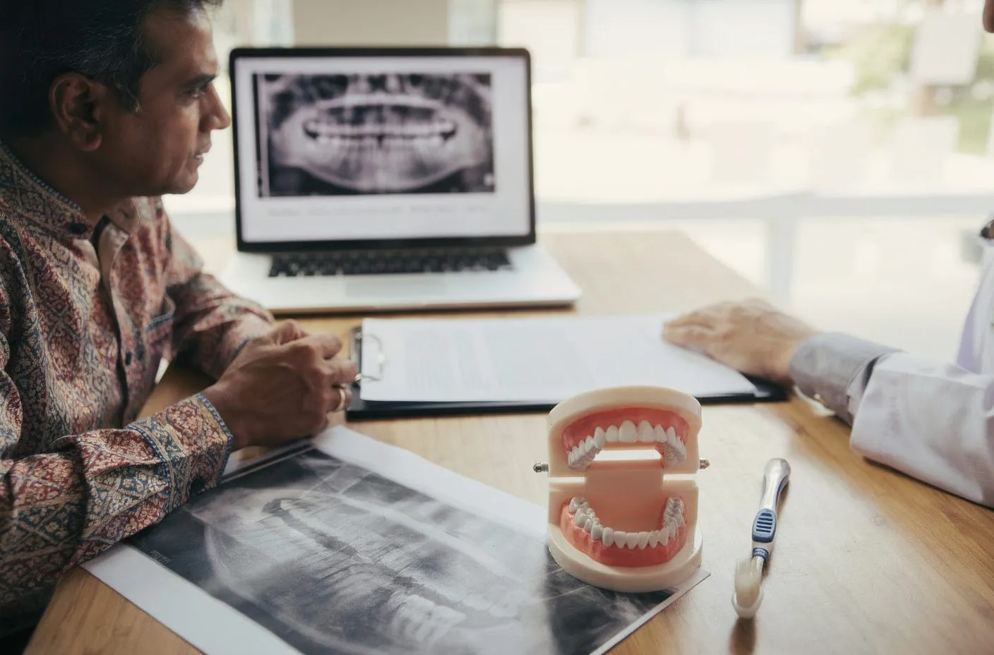
"Every doctor I have been to has shown me a film X-ray and given me a different answer," he said. "One said save it, one said remove it, one said wait and see. I want someone to actually show me what is happening inside."
A CBCT scan gave us the answer in under 10 minutes. The original root canal had missed a fourth canal in the mesiobuccal root — a known anatomical variant in upper first molars. The untreated canal had a small periapical lesion around it, which explained the recurring symptoms. The remaining 3 root fills were intact and well-condensed. The tooth itself was structurally sound, with adequate bone support and no vertical fracture.
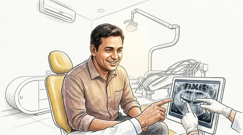
I walked Ashish through the CBCT cross-sections on screen. "This is exactly what you asked for — here is what is happening inside. The original treatment missed this canal. It is accessible under the microscope. Retreatment success for a case like this runs at around 80 to 85%. If retreatment fails, we extract and place an implant — implant survival at 5 years is 98%. But the evidence favours trying to save the tooth first."
Ashish agreed to microscope-assisted modern dentistry retreatment. Under the operating microscope, the MB2 canal was located, negotiated, and cleaned to length. The full procedure took 90 minutes. The tooth was temporised and we reviewed at 3 months.
"No ache at all since the week after the treatment," he reported at the 3-month appointment. The CBCT review showed the periapical lesion had reduced by approximately 60%. The tooth was restored with a full-coverage ceramic crown designed using an intra-oral scan — no putty impression, a single scan that took 4 minutes.
At the twelve-month review, the lesion had resolved completely on CBCT. The crown was intact, the bite was comfortable, and Ashish had recommended the clinic to 3 colleagues who had also been told they needed implants without adequate imaging to support the diagnosis.
"The difference was being shown the problem rather than just being told about it," he said. "When I could see the scan myself, the decision was straightforward."
BDS, MDS Prosthodontist · DCI A-04860 · Smile Gallery, Bhopal
| Follow-up | 12 months post-retreatment |
| Periapical lesion | Fully resolved on CBCT at 12 months |
| Tooth retained | Yes — no extraction required |
| Restoration | Full-coverage ceramic crown via intra-oral scan |
| Ongoing care | 6-monthly check-up, annual CBCT if symptomatic |
Frequently Asked Questions
What modern technologies are available in Bhopal’s dental clinics?
Digital X-rays, intra-oral scanners, CBCT imaging, laser-assisted procedures, CAD/CAM and 3D-printed restorations, microscope-assisted endodontics, and digital smile design.
Are these technologies available at Smile Gallery in Bhopal?
Yes. Smile Gallery, in Arera Colony, uses these tools in routine practice under Dr. Saurabh Shrivastava (DCI: A-04860).
How long does a typical visit using digital tools take?
A digital scan replaces a 10-minute impression with a 5-minute scan. Crown delivery visits run 30 to 45 minutes.
What should I expect at my first visit?
An unhurried consultation, X-rays viewed on screen, photographs where relevant, and a written treatment plan.
How do I book an appointment at Smile Gallery, Arera Colony?
Call +91 9200700750.
Ready for a consultation?
Visit Smile Gallery Dental Wellness Centre, E-4/205, Main Rd 3, near Flower Market, E-4, Arera Colony, Bhopal.
Open Monday to Saturday 10am–2pm and 5–9pm.
