Dental X-rays let your dentist see decay between teeth, infection at tooth roots, bone loss, impacted wisdom teeth, and developing problems before they cause pain. Modern digital X-rays use very low radiation and are an essential part of accurate dental treatment planning.
Medically reviewed by Dr. Saurabh Shrivastava, BDS MDS Prosthodontist, Certified Digital Smile Designer (DSD) (DCI: A-04860). Last updated: May 2026.
Dental X-rays let your dentist see what the eye cannot — decay between teeth, infection at the root tip, hidden bone loss, impacted wisdom teeth, and the precise position of structures inside the jaw — and that visibility is what allows Dr. Saurabh Shrivastava, BDS MDS Prosthodontist (DCI: A-04860) at Smile Gallery Dental Wellness Centre, Arera Colony, to plan accurate dental treatment for patients from Awadhpuri and surrounding areas. When a deep cavity is caught early on an X-ray, root canal treatment may be avoided altogether; even when it is needed, root canal therapy has a 95% success rate over 10 years.
Why X-rays Are Essential to Modern Dentistry
A clinical exam shows the visible surfaces of teeth and the gum line — but a dental X-ray reveals what lies beneath. Many problems start in places the eye cannot reach: between adjacent teeth where contact points hide cavities, beneath fillings or crowns, inside the root canal system, and within the bone that supports the teeth. X-rays make these regions visible and allow problems to be treated when they are still small and simple to fix.
Types of Dental X-rays You May Encounter
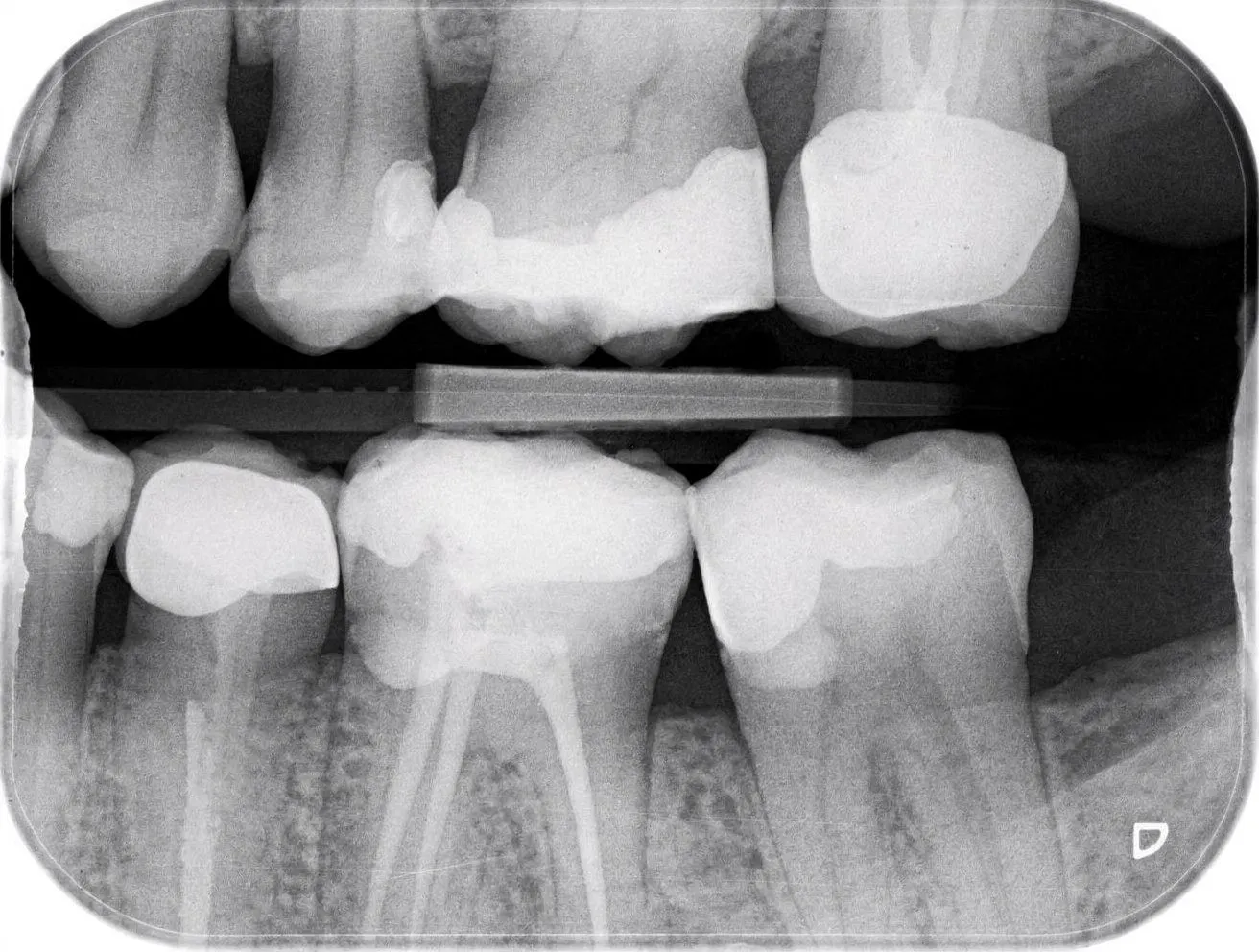
Bitewing X-rays show the upper and lower back teeth on one side and are excellent for catching cavities between teeth. Periapical X-rays show a single tooth from crown to root tip and are used for diagnosing infection or planning root canal treatment. Panoramic X-rays show the entire jaw in a single image and are useful for wisdom teeth, full-mouth assessment, and orthodontic planning. CBCT scans give a three-dimensional view, especially useful for implant planning and complex cases.
According to Dr. Saurabh Shrivastava, MDS Prosthodontist: "A clinical exam shows me the top of the tooth — it does not show me what is happening between teeth, under fillings, or at the root tip inside the bone. A bitewing X-ray taken at the right time catches the cavity that would otherwise require a root canal six months later. That is a significantly different outcome for the patient."
How Modern Digital X-rays Work
Digital sensors have replaced film for most everyday X-rays. They produce sharper images, allow magnification on screen, and use significantly less radiation than older film systems. The image appears within seconds, so the dentist can show you the finding and explain it during the same visit — a small but real shift in how care is delivered.
"The radiation dose from a modern digital dental X-ray is comparable to a few hours of natural background radiation — less than a short flight. The risk of not taking the X-ray, on the other hand, is a missed diagnosis that turns a small cavity into a root canal, or an early infection into an extraction. That is not a difficult calculation."
Dr. Saurabh Shrivastava · BDS, MDS Prosthodontist, DCI A-04860
How Often Are X-rays Needed?
For a healthy adult with no recent cavities, bitewing X-rays every one to two years are typical. For someone with multiple recent fillings, active gum disease, or a history of cavities, more frequent imaging may be sensible. For a new patient, an initial set of X-rays establishes a baseline. Children usually need fewer X-rays unless decay is suspected or orthodontic planning is under way.
- Bitewing X-ray — shows the crowns of upper and lower back teeth on one side simultaneously. The primary tool for catching cavities between teeth (interproximal decay) before they are clinically visible. Recommended every 1 to 2 years for adults with no active decay.
- Periapical X-ray — shows a single tooth from the biting edge to the tip of the root and the surrounding bone. Used to diagnose infection at the root tip, monitor root canal treatment, plan extractions, and assess individual tooth health.
- Panoramic X-ray (OPG) — shows the entire upper and lower jaw, all teeth, both joints, and the sinus floors in a single image. Used for new patient assessment, wisdom tooth evaluation, orthodontic planning, and full-mouth overview.
- CBCT scan — a three-dimensional cone beam computed tomography scan showing bone volume, nerve location, sinus anatomy, and root morphology. Essential for implant planning, complex extractions, and any case where two-dimensional imaging is insufficient.
- Intra-oral periapical series — a set of periapical X-rays covering the full mouth, usually 14 to 18 films. Used for comprehensive new patient records, periodontal disease staging, and cases requiring detailed individual tooth assessment across the entire dentition.
Are Dental X-rays Safe?
Yes, when used appropriately. The radiation dose from a modern digital dental X-ray is very low — comparable to a few hours of natural background radiation. Lead aprons and thyroid collars further protect patients. For pregnant patients, non-essential imaging is deferred where possible, and emergency imaging is shielded carefully.
According to Dr. Saurabh Shrivastava, MDS Prosthodontist: "For implant planning we use CBCT — a three-dimensional scan that shows bone height, width, and the precise location of the inferior alveolar nerve. Without it, placing an implant is guesswork with someone's jaw. With it, I can plan the exact position, angle, and depth on screen before the patient sits in the chair."

The clinical case and outcome are from Dr. Saurabh Shrivastava's practice.
Suresh came in on a Monday morning in September, recently retired from a government department in Habibganj and finally, as he put it, "getting around to things I kept putting off." He was 52, had not seen a dentist in 8 years, and had no specific complaint. "I just want to know how things stand," he said, which is exactly the right reason to come.
The clinical examination showed a full set of teeth, healthy-looking gum colour, and no obvious cavities on direct inspection. His oral hygiene was reasonable — light calculus on the lower anterior lingual surfaces but nothing severe. He had a large amalgam filling in the upper left second molar that appeared intact. Based on visual examination alone, I might have given him a reasonable report.
The bitewing X-rays changed the picture. Two early cavities between the upper right premolars — classic interproximal decay, invisible clinically and invisible to Suresh. More importantly, the panoramic X-ray showed a periapical radiolucency at the root tip of the upper left second molar — a shadow indicating infection at the end of the root, almost certainly related to the old amalgam filling, which had likely been leaking for years. Suresh had no pain and no symptoms. He would have gone home with a clean bill of health and an infection that would have eventually abscess in the most inconvenient moment possible.
I showed him the X-ray on screen and pointed to the shadow. "This grey area around the root tip is infection in the bone. It has been there long enough to create this size of lesion — probably 2 to 3 years. Because it is not pressing on a nerve, you feel nothing. But left alone, it will continue to expand and eventually become a painful abscess or require extraction."
"And now that you've found it?" he asked.
"Root canal treatment — 2 visits. We clean the canal, seal it, and the infection resolves over 3 to 6 months as the bone fills back in. The dental X-ray at your six-month follow-up will show the lesion shrinking. The 2 small cavities between the premolars can be filled in the same first appointment."
We completed the root canal in 2 appointments — the first cleaning and shaping, the second obturation and a temporary seal. A new porcelain crown was placed over the molar 2 weeks after the second appointment to protect the root-canal-treated tooth. The 2 premolar cavities were filled with composite resin in the first appointment — 20 minutes for both.
At the 6-month follow-up X-ray, the periapical lesion had reduced noticeably — the bone shadow was smaller and its edges were becoming less defined, indicating new bone deposition. Suresh was asymptomatic throughout. The crown was intact, the composite fillings were sealed.
"Eight years without checking," he said, looking at the comparison X-rays. "I didn't think anything was happening because nothing hurt. I'll be back every six months now." He booked the next appointment before leaving — and brought his wife in the following week for her first check-up in five years.
BDS, MDS Prosthodontist · DCI A-04860 · Smile Gallery, Bhopal
| Follow-up | 6 months post-root canal |
| Periapical lesion | Reducing on X-ray — bone deposition visible at 6 months |
| Crown | Porcelain crown intact, no sensitivity |
| Premolar fillings | 2 composite fillings sealed, no secondary decay |
| Ongoing care | 6-monthly check-up with bitewing X-ray at alternating visits |
Frequently Asked Questions
Why does my dentist want X-rays at the first visit?
To establish a baseline view of teeth and bone, and to detect any hidden decay or infection that should be treated early.
Are X-rays available at Smile Gallery in Bhopal?
Yes. Smile Gallery, in Arera Colony, uses digital X-rays and CBCT imaging where indicated, with results shown to patients during the same visit.
How long does a dental X-ray take?
A bitewing or periapical X-ray takes a few seconds. A panoramic X-ray takes around 15 to 20 seconds. A CBCT scan takes a similar amount of time.
What should I expect after the X-ray?
Nothing — there is no recovery. The dentist reviews the image with you on screen and explains any findings.
How do I book an appointment at Smile Gallery, Arera Colony?
Call +91 9200700750. Bring any previous X-rays you have so they can be compared.
Ready for a consultation?
Visit Smile Gallery Dental Wellness Centre, E-4/205, Main Rd 3, near Flower Market, E-4, Arera Colony, Bhopal.
Open Monday to Saturday 10am–2pm and 5–9pm.
